The brain is a complex organ that generates electrical activity. By measuring these electrical signals, healthcare professionals can gain valuable insights into brain function and diagnose various neurological conditions. One of the most common methods used to test brain waves is through an Electroencephalogram (EEG). In this article, we will explore how an EEG test works, its applications, and what to expect during the procedure.
Using an EEG test
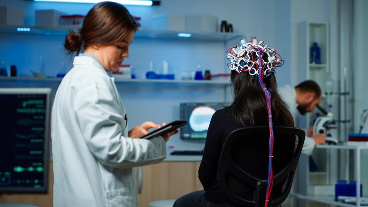
An EEG test involves the application of electrodes to the scalp to detect and record the electrical activity in the brain. These electrodes are connected to an amplifier, which amplifies the electrical charges and displays them as a graph on a computer screen or prints them out on paper.
Applying electrodes to the scalp
Before the EEG test begins, the healthcare provider will clean the scalp and apply a conductive gel or paste to ensure good contact between the electrodes and the skin. The electrodes are then attached to specific locations on the scalp using adhesive or a cap.
Amplifying and displaying the electrical charges
Once the electrodes are in place, the electrical charges generated by the brain are picked up by the electrodes and sent to an amplifier. The amplifier increases the strength of the electrical signals, making them easier to analyze. The amplified signals are then displayed as a graph on a computer screen or printed out on paper.
Interpreting the readings
The healthcare provider will analyze the EEG readings to identify patterns and abnormalities in the brain waves. Different brain wave patterns are associated with different states of consciousness, such as wakefulness, sleep, and certain neurological conditions. By interpreting the readings, healthcare professionals can diagnose conditions like epilepsy, tumors, stroke, Alzheimer’s disease, psychoses, narcolepsy, and coma.
Conducting evoked potential studies
In addition to measuring ongoing brain activity, an EEG can also be used to conduct evoked potential studies. These studies involve stimulating the brain with specific sensory inputs, such as flashing lights or auditory tones, and measuring the brain’s response. This can provide valuable information about the integrity of the sensory pathways and help diagnose conditions like multiple sclerosis or nerve damage.
Measuring brain activity in response to stimulation
During an evoked potential study, the healthcare provider will present the stimuli and measure the brain’s response using the EEG. The brain’s response is typically seen as a change in the pattern of the brain waves. By analyzing these changes, healthcare professionals can assess the function of specific sensory pathways and identify any abnormalities.
Identifying abnormalities in brain waves
One of the main purposes of an EEG is to identify abnormalities in brain wave patterns. Certain conditions can cause the brain waves to deviate from the normal patterns, indicating underlying neurological issues.
Rapid spiking waves or slow waves
Rapid spiking waves, also known as epileptiform discharges, are abnormal electrical patterns that can indicate epilepsy or seizure activity. Slow waves, on the other hand, may be seen in conditions like dementia or brain damage. By identifying these abnormal patterns, healthcare professionals can make accurate diagnoses and develop appropriate treatment plans.
Diagnosing brain disorders
EEG is a valuable tool for diagnosing various brain disorders. By analyzing the brain wave patterns, healthcare professionals can identify abnormalities that are indicative of specific conditions.
Epilepsy, tumors, stroke, Alzheimer’s disease, psychoses, narcolepsy, coma
EEG is commonly used to diagnose epilepsy, a neurological disorder characterized by recurrent seizures. The presence of abnormal electrical activity in the brain can confirm the diagnosis and help determine the type of epilepsy and the most effective treatment options.
In addition to epilepsy, an EEG can also help diagnose other conditions such as brain tumors, stroke, Alzheimer’s disease, psychoses, narcolepsy, and coma. Each of these conditions has distinct EEG patterns that can aid in their diagnosis.
Monitoring blood flow during surgery
EEG can also be used to monitor blood flow in the brain or neck’s blood vessels during surgery. By measuring the electrical activity in the brain, healthcare professionals can assess the adequacy of blood supply and detect any abnormalities that may require intervention.
In the brain or neck’s blood vessels
During surgery, electrodes are placed on the scalp or neck to monitor the electrical activity in the brain or blood vessels. Any changes in the EEG readings can alert the surgical team to potential issues with blood flow, allowing them to take appropriate action to ensure the patient’s safety.
Risks and factors to consider
While an EEG is generally considered safe, there are some risks and factors that need to be taken into consideration.
Risks of seizures during the test
Although rare, there is a small risk of inducing a seizure during an EEG test, especially in individuals with a history of epilepsy. Healthcare providers are trained to recognize and manage seizures if they occur during the procedure.
Factors that may interfere with the EEG reading
Several factors can interfere with the accuracy of the EEG reading. These include low blood sugar, body or eye movement, bright or flashing lights, certain medications, and caffeine. It is important to inform the healthcare provider about any medications or substances that may affect the EEG results.
Preparing for the EEG
Before undergoing an EEG, there are certain preparations that need to be made to ensure accurate results.
Washing hair and avoiding hair styling products
Prior to the test, it is important to wash the hair thoroughly and avoid using any hair styling products, such as gels, sprays, or oils. This helps to ensure good contact between the electrodes and the scalp.
Informing healthcare provider about medications and supplements
It is crucial to inform the healthcare provider about all medications, including over-the-counter drugs and herbal supplements, being taken. Some medications can interfere with the EEG readings and may need to be temporarily discontinued before the test.
Following fasting and sleep instructions
Depending on the purpose of the EEG, fasting or sleep instructions may need to be followed. For example, if the EEG is being done to evaluate sleep disorders, the patient may be asked to sleep for a specific number of hours before the test.
During the EEG
During the EEG test, the patient will be comfortably positioned in a reclining chair or lying on a bed.
Attaching electrodes to the scalp
The healthcare provider will attach the electrodes to specific locations on the scalp using a special paste or a cap. The number and placement of the electrodes may vary depending on the purpose of the EEG.
Remaining still and avoiding movements
It is important for the patient to remain as still as possible during the EEG to ensure accurate readings. Any movements, such as body or eye movements, can interfere with the electrical signals and affect the results.
Undergoing additional tests with stimuli
In some cases, the healthcare provider may administer additional tests during the EEG, such as deep breathing or flashing lights. These tests are designed to elicit specific responses in the brain and provide further information about its function.
After the EEG
After the EEG test is complete, there are a few things to keep in mind.
Expected duration of the EEG
An EEG typically takes between 45 minutes to 2 hours to complete, depending on the specific requirements of the test.
Prolonged EEG monitoring or ambulatory EEG
In some cases, a prolonged EEG monitoring or ambulatory EEG may be required. These tests involve wearing the electrodes for an extended period, usually 24 hours or more, to capture brain activity over a longer period of time.
Removing electrodes and washing off electrode paste
After the test, the healthcare provider will remove the electrodes from the scalp. Any residual electrode paste can be easily washed off with warm water and mild soap.
Resting and arranging transportation if sedatives were used
If sedatives were used during the EEG, it is important to rest and avoid any activities that require alertness. It is also advisable to arrange for someone to drive you home, as sedatives can impair coordination and judgment.
Possible skin irritation or redness at electrode locations
It is common to experience mild skin irritation or redness at the electrode locations. This usually subsides within a few hours or days and can be relieved with over-the-counter creams or ointments.
Following additional instructions from healthcare provider
After the EEG, the healthcare provider may provide additional instructions or discuss the results of the test. It is important to follow these instructions and schedule any necessary follow-up appointments.